Datei:Axial HRUS neck scans & vagus cs measurements Walter et al (2018) FrontNeurol 9-805.jpg
Zur Navigation springen
Zur Suche springen

Größe dieser Vorschau: 426 × 599 Pixel. Weitere Auflösungen: 170 × 240 Pixel | 341 × 480 Pixel | 546 × 768 Pixel | 1.099 × 1.546 Pixel
Originaldatei (1.099 × 1.546 Pixel, Dateigröße: 510 KB, MIME-Typ: image/jpeg)
![]()
Diese Datei und die Informationen unter dem roten Trennstrich werden aus dem zentralen Medienarchiv Wikimedia Commons eingebunden.
![]()
_FrontNeurol_9-805.jpg?uselang=de)
Beschreibung
| Beschreibung |
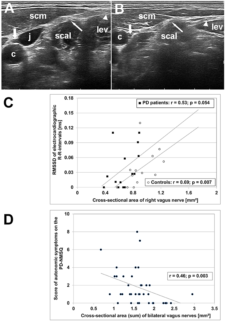
English: Original figure caption: High-resolution ultrasonography (HR-US) findings of vagus, spinal accessory and phrenic nerves in PD patients and controls. (A) Axial HR-US scan of the lateral cervical region at the midneck level in a healthy elderly woman. The vagus nerve (thick arrow) is visualized in the carotid sheath between the common carotid artery (c) and the jugular vein (j). The phrenic nerve (thin arrow) is located superficial to the scalene muscle (scal) underneath the sternocleidomastoid muscle (scm). The spinal accessory nerve (triangle) is identified superficial to the levator scapulae muscle (lev). (B) Axial HR-US scan of the lateral cervical region in a PD patient. Compared to the control subject shown in (A) the vagus nerve (thick arrow) shows a clearly reduced caliber. The phrenic nerve (thin arrow) and the spinal accessory nerve (triangle) are of similar size as in age-matched controls. (C) Diagram showing the correlation between RMSSD, an electrocardiographic parameter reflecting vagal cardiac innervation, and caliber of right vagus nerve in PD patients (°) and age-matched controls (■). (D) Diagram showing the correlation between the sum score of autonomic symptoms on the PD Non-Motor Symptoms Questionnaire (calculated from items 1, 3, 4, 5, 6, 7, 8, 11, 19, 20, 28) and bilateral vagus nerve caliber in the combined group of PD patients and age-matched controls. Deutsch: Befunde hochauflösender Sonographie (engl. high-resolution ultrasonography, HRUS) des Nervus vagus, N. accessorius und N. phrenicus bei Parkinson-Patienten und Kontrollpersonen. (A) Axialer HRUS-Scan der lateralen Halsregion auf mittlerer Höhe bei einer gesunden älteren Frau. Der Vagusnerv (fetter Pfeil) ist erkennbar in der Carotisscheide zwischen der Carotis communis (c) und der Jugularis interna (j). Der Phrenicusnerv (dünner Pfeil) befindet sich an der Oberfläche des Musculus scalenus anterior (scal) unterhalb des M. sternocleidomastoideus (scm). Der Accessoriusnerv (Dreieck) ist an der Oberfläche des des M. levator scapulae (lev) zu erkennen. (B) Axialer HRUS-Scan der lateralen Halsregion bei einem Parkinson-Patient. Im Vergleich zur in (A) gezeigten Kontrollperson zeigt der Vagusnerv eine deutlich geringere Querschnittsfläche. Der Phrenicus- (dünner Pfeil) und Accessoriusnerv (Dreieck) haben die gleiche Größe wie bei gleichaltrigen Kontrollpersonen. (C) Diagramm mit Korrelation zwischen RMSSD, einem elektrokardiographischen Parameter, der die Innervation des Herzens durch den Vagus abbildet, und der Querschnittsfläche des rechten Vagusnervs bei Parkinson-Patienten (°) und gleichaltrigen Kontrollpersonen (■). (D) Diagramm mit Korrelation der Punktzahl bei autonomen Symptomen des PD Non-Motor Symptoms Questionnaire (ein Fragebogen zur qualitatativen Erfassung der nicht-motorischen Störungen bei Parkinson-Patienten; Punktzahl errechnet aus den Items 1, 3, 4, 5, 6, 7, 8, 11, 19, 20, 28) und der beidseitigen Querschnittsfläche des Vagusnervs in der Mischgruppe aus Parkinson-Patienten und Kontrollpersonen. |
| Datum | (publication date) |
| Quelle | Fig. 1 in: Atrophy of the Vagus Nerve in Parkinson’s Disease revealed by High-Resolution Ultrasonography. Frontiers in Neurology 9:805, doi:10.3389/fneur.2018.00805 |
| Urheber | Uwe Walter, Panagiota Tsiberidou, Maxi Kersten, Alexander Storch, Matthias Löhle |
| Genehmigung (Weiternutzung dieser Datei) |
This image was published in Frontiers in Neurology journal. On the website that contains the HTML version of the respective article (see DOI link above) it is stated that it “is an open-access article distributed under the terms of the Creative Commons Attribution License (CC BY). The use, distribution or reproduction in other forums is permitted, provided the original author(s) or licensor are credited and that the original publication in this journal is cited, in accordance with accepted academic practice.” |
Lizenz
Diese Datei ist lizenziert unter der Creative-Commons-Lizenz „Namensnennung 4.0 international“.
- Dieses Werk darf von dir
- verbreitet werden – vervielfältigt, verbreitet und öffentlich zugänglich gemacht werden
- neu zusammengestellt werden – abgewandelt und bearbeitet werden
- Zu den folgenden Bedingungen:
- Namensnennung – Du musst angemessene Urheber- und Rechteangaben machen, einen Link zur Lizenz beifügen und angeben, ob Änderungen vorgenommen wurden. Diese Angaben dürfen in jeder angemessenen Art und Weise gemacht werden, allerdings nicht so, dass der Eindruck entsteht, der Lizenzgeber unterstütze gerade dich oder deine Nutzung besonders.
Dateiversionen
Klicke auf einen Zeitpunkt, um diese Version zu laden.
| Version vom | Vorschaubild | Maße | Benutzer | Kommentar | |
|---|---|---|---|---|---|
| aktuell | 03:22, 14. Apr. 2019 | | 1.099 × 1.546 (510 KB) | Gretarsson | {{Information |description ={{en|1=Original figure caption: ''High-resolution ultrasonography (HR-US) findings of vagus, spinal accessory and phrenic nerves in PD patients and controls. (A) Axial HR-US scan of the lateral cervical region at the midneck level in a healthy elderly woman. The vagus nerve (thick arrow) is visualized in the carotid sheath between the common carotid artery (c) and the jugular vein (j). The phrenic nerve (thin arrow) is located superficial to the scalene muscle (s... |
Dateiverwendung
Die folgende Seite verwendet diese Datei:
_FrontNeurol_9-805.jpg)