Zur Beschreibungsseite auf Commons

Datei:Electron micrograph of neuromuscular junction (cross-section).jpg

aus Wikipedia, der freien Enzyklopädie
Zur Navigation springen Zur Suche springen

Electron_micrograph_of_neuromuscular_junction_(cross-section).jpg(433 × 289 Pixel, Dateigröße: 95 KB, MIME-Typ: image/jpeg)

Diese Datei und die Informationen unter dem roten Trennstrich werden aus dem zentralen Medienarchiv Wikimedia Commons eingebunden.

Zur Beschreibungsseite auf Commons


Beschreibung

Beschreibung
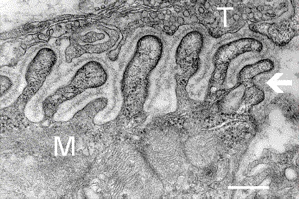
English: Electron micrograph showing a cross-section through the neuromuscular junction. T is the axon terminal, M is the muscle fiber. The arrow shows junctional folds with basal lamina. Postsynaptic densities are visible on the tips between the folds. The scale is 0.3 µm.
Datum Originally uploaded to en.wikipedia on 10. März 2006.
Quelle Synapse Web at the National Institute of Mental Health, National Institutes of Health; originally from en.wikipedia; description page is/was here.
Urheber National Institute of Mental Health; originally uploaded by Nrets at en.wikipedia.

Lizenz

Gemeinfrei Dieses Bild ist ein Werk des National Institutes of Health, Teil des Ministeriums für Gesundheitspflege und Soziale Dienste der Vereinigten Staaten, aufgenommen oder erstellt als Teil der offiziellen Pflichten eines Mitarbeitenden. Als Werk der Bundesregierung der Vereinigte Staaten ist das Bild in der Gemeinfreiheit.
Bitte stelle sicher, dass dieses Bild tatsächlich von der US-Bundesregierung erstellt wurde. Das NIH verwenden häufig kommerzielle Bilder, die nicht gemeinfrei sind.


Ursprüngliches Datei-Logbuch

(All user names refer to en.wikipedia)

  • 2006-03-10 20:07 Nrets 433×289×8 (97758 bytes) Electron micrograph showing a cross section through the neuromuscular junction. T is the axon terminal, M is the muscle fiber. The arrow shows junctional folds with basal lamina. Postsynaptic densities are visible on the tips between the folds. Scale is 0

Kurzbeschreibungen

Ergänze eine einzeilige Erklärung, was diese Datei darstellt.

In dieser Datei abgebildete Objekte

Motiv

Dateiversionen

Klicke auf einen Zeitpunkt, um diese Version zu laden.

Version vomVorschaubildMaßeBenutzerKommentar
aktuell05:41, 22. Mär. 2007Vorschaubild der Version vom 05:41, 22. Mär. 2007433 × 289 (95 KB)Fran Rogers{{Information |Description=Electron micrograph showing a cross section through the neuromuscular junction. T is the axon terminal, M is the muscle fiber. The arrow shows junctional folds with basal lamina. Postsynaptic densities are visible on the tips be

Die folgende Seite verwendet diese Datei:

Globale Dateiverwendung

Die nachfolgenden anderen Wikis verwenden diese Datei:

Metadaten